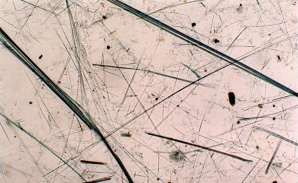
The identification of asbestos minerals is based on:
- the appearance of the minerals: long fibers (length:thickness > 3) forming bundles; fibers split into fine fibrils
- the chemical composition of the minerals
- the fact that the material is crystalline
The asbestos minerals can be identified using several methods. Commonly, phase contrast polarized light microscopy (PLM) and transmission electron microscopy (TEM) are used. Other potential techniques include X-ray diffraction (XRD) and possibly infra-red spectroscopy (IRS).
Scanning electron microscopy (SEM)
Scanning electron microscopy is primarily used for asbestos analyses in air and dust samples. The asbestos fibers are identified using their
- chemical composition
- appearance
With SEM, asbestos fibers too fine for detection with light microscopy can be detected.
Analyses for asbestos in dust are usually performed in order to assess if asbestos has been dispersed during asbestos removal work.
Likewise, the asbestos concentration in air is tested after removal work to ascertain that these hazardous fibers have not spread to the surroundings.
Phase contrast polarizied light microscopy (PLM)
Asbestos fibers can be identified using
- appearance
- optical properties
- refractive index
The presence of asbestos in a material sample is normally confirmed by using PLM. The asbestos minerals can be identified by their appearance in a microscope. They form bundles, and the individual fibers split at the ends. The fibers have to be crystalline, and the appearance has to be in accordance with the optical properties of the asbestos minerals.
The asbestos minerals have characteristic refractive indices. By embedding the fibers in well-defined index-matching liquids, the different asbestos types can be distinguished. Phase contrast microscopy is also used for fiber counting.
